Closing the In Vitro–In Vivo Gap: Organ-on-Chip, Imaging, and PBPK Integration for Mesoporous Materials, Oral/Targeted Nanomedicine and Advanced Delivery
Md. Humayan Kabir (Master of Science in Pharmaceutical Technology)
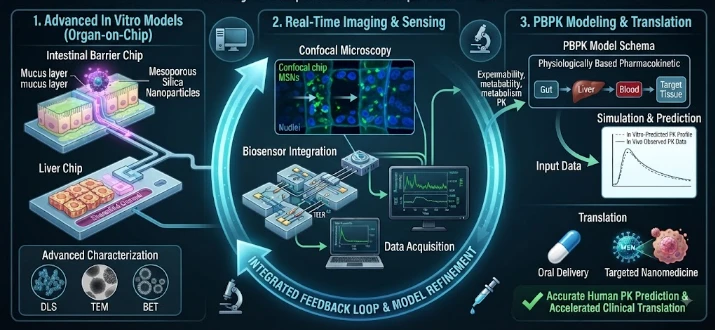
This article proposes an integrated strategy for closing the in vitro–in vivo gap in mesoporous drug-delivery systems using organ-on-chip models, imaging, and PBPK modeling. It explains that attractive laboratory results for dissolution, uptake, or targeted delivery may fail in vivo because biological fluids, mucus, epithelial barriers, vascular flow, protein corona formation, immune clearance, degradation, and tissue architecture change nanoparticle behavior. For oral delivery, gut models can reproduce flow, mucus, epithelial transport, pH, and local retention; for systemic delivery, vascularized chips can test adhesion, translocation, and uptake under flow. Imaging provides spatial information, but the article stresses that tracer stability and chemical validation are essential because labels may persist after the carrier degrades. PBPK models then connect local transport measurements to whole-body concentration–time behavior, ideally distinguishing intact carrier, released drug, degraded material, and metabolites. Sensitivity analysis guides the next experiment, while disease-specific models, repeated dosing, realistic concentrations, batch variation, and falsifiable mechanistic predictions improve translation. The approach treats material properties, biological transformation, tissue exposure, and whole-body fate as one connected problem.
The unique benefits of mesoporous silica and other porous nanomaterials, such as high drug loading, pore parameters that are easily engineered, surface modification and tunable release, make them an attractive choice for drug delivery applications. Laboratory studies consistently demonstrate enhanced dissolution, targeted uptake or stimuli-responsive behavior. Translation is less predictable in that a particle enters into a complex biological environment where particle identity on the surface is altered, transported, degraded, and cleared. These complementary tools allow for the connection of attractive in vitro performance with realistic in vivo exposure: organ-on-chip systems, imaging and physiologically based pharmacokinetic modeling.
The starting point to the gap is the in vitro model. The release of the drug in a simple buffer does not mean much for the drug release in mucus, proteins, enzymes, changing pH, passage through the intestine, vascular flow and tissue barriers. Static cell monolayers only provide biology but are missing key mechanical and spatial characteristics. Therefore, the testing environments need to have similar characteristics to the route and therapeutic target of mesoporous materials.
In the case of oral delivery, dissolution, gastrointestinal fluids, mucus diffusion, epithelial transport, particle degradation and local retention must be taken into consideration for a realistic model. Mesoporous Silica may interact with inflamed and healthy gastrointestinal tissue differently due to surface chemistry. Microfluidic gut models provide flow, organization of the epithelia, and regulated luminal conditions. These systems can detect if there is improved drug transport due to actual transport benefits or due to artifacts from static culture.
But, targeted systemic delivery presents another challenge. Once injected into the bloodstream, nanoparticles undergo quick interactions with plasma proteins and cells, forming a biomolecular corona that may alter the nanoparticles' targeting behavior. Competition of endothelial barriers, macrophages, liver sinusoids, spleen filtration, and tumor vasculature follow. Selected aspects of these processes can be recapitulated using the vascularized organ-on-chip model and can be directly observed under flow conditions to analyze adhesion, translocation and cellular uptake.
The properties of particles should be measurable during these experiments. Changes in size, aspect ratio, pore structure, surface charge, coating stability, degradation and payload release can occur in biological media. Only a partial picture is given from a carrier who is only characterized prior to exposure. Determination of the biological behaviour observed either through recovery of particles or the in situ analytical methods, can be used to distinguish between the original biological behaviour and transformational behaviour.
Imaging - the spatial bridge. Fluorescence microscopy is used to monitor localization of cells or tissue chips and confocal or high-content imaging to resolve intracellular distribution. Depending on the material design, in animal studies, the accumulation of the material can be estimated by using fluorescence, positron emission tomography, magnetic resonance or other modalities. When fluorescent labeling can be a concern for loose attachment to carrier, the elemental analysis option is available to add quantitative silicon measurements.
Special attention must be paid to the stability of the labels. When the pore structure is degraded, leaving behind a signal may give the appearance of persistence of the intact particles. Studies with variously labeled radionuclides have shown that degradation and biodistribution interpretation depends on the position of the tracer. A chemical validation of the imaging is then required, to avoid the misidentification of the signal location with the location of the functional carrier.
Those spatial observations are transformed to a kinetic system using PBPK modeling. Compartments are blood and relevant organs; parameters are flow, tissue uptake, release, degradation, metabolism of payload and elimination. There is a need for extra structure in oral systems for absorption and gastrointestinal transit. The model links between local experimental and whole body concentration-time predictions.
In conventional PBPK assumptions, only a single species exists and needs to be accounted for.Multiple species exist and need to be accounted for in Nanomaterials, which complicates conventional PBPK assumptions. The different routes can be taken by intact carrier, partially degraded particle, released drug, metabolite, and detached surface components. Analytical data, when available, are most appropriate for multi-species models which better represent this reality. A simplified particle-bound/free drug distinction can even significantly enhance the mechanistic interpretation.
Parameters that can be provided by organ-on-chip measurements. The permeability to the barrier, the uptake rate, the intracellular retention, the flow assisted transport, and the release kinetics can be determined experimentally. Parameter uncertainty is then fed into the PBPK model. Sensitivity analysis is used to determine which biological process has the biggest influence on the target exposure predicted by the chip experiments, and then to determine the subsequent experiments to conduct.
This forms a circularity. The chip indicates a transport rate, the PBPK model calculates an organ exposure, imaging tests verify the prediction in vivo, and discrepancies lead to model or experiment improvement. It's the structured contradiction that's important. Rather than animal biodistribution being a one-off confirmation procedure, unexpected results indicate that the assumption of the mechanism was wrong.
Translation is further enhanced by disease-specific models. Altered mucus and permeability in inflamed intestine, changes in vascular architecture of tumors, changes in diffusion in fibrotic tissue and liver disease changing clearance. Designed cells or chips that mimic the disease mimic certain features of the patient. PBPK population simulations are then used to investigate the impact of these differences on exposure over a range of clinical phenotypes.
This is especially advantageous for mesoporous oral formulations as apparent bioavailability improvements may occur due to multiple mechanisms. It may be due to increased dissolution rates in the drug, supersaturation, shift in precipitation, change in the uptake of the carrier, extended local residence or permeability changes. These mechanisms can be separated for prediction of the performance across food states, GI pH variation and disease.
This is a similar problem with targeted nanomedicine. The high fluorescence of the tumor does not guarantee receptor-mediated targeting since the same appearance may be obtained by vascular permeability or by uptake by macrophages or by release of the label. Competition studies, receptor-negative controls, quantitative imaging and chip-based transport assays elucidate mechanism. PBPK models are then used to determine if the targeting contribution is significant in comparison to the distribution.
Integration also enhances safety. Organ-specific chips and in vivo imaging can be used to identify liver or spleen accumulation, slow degradation, inflammatory signaling and persistent tissue exposure. A PBPK simulation accurately predict the changes in accumulation with repeated dosing. Safety margins are no longer only related to the nominal dose administered, but to tissue burden.
The characterization of the protein-crowns is another pathway that connects chips to in vivo behavior. Analysis of the proteins that are adsorbed can provide information on whether the surface functionalization alters the opsonisation, complement-related interactions or recognition through the phagocytes. Chips with endothelium and/or macrophage populations are then used to study any functional consequences of flow. Parameters of PBPK models for uptake or clearance become more mechanistic in nature, rather than being fitted as unexplained empirical constants.
It is also important that barrier models have realistic concentration ranges. Studies of nanoparticles tend to involve high in vitro doses that are more than likely to be delivered to tissue following administration. PBPK simulations are also able to go in the direction that predicts concentrations that may occur at a target organ. Experiments performed in those ranges yield more useful permeability and toxicity parameters for the chip experiments, and establish a closer connection between the modeling and the experiment design.
The term "batch variation" should be specifically challenged. Two mesoporous preparations having the same average pore size may have different accessibility of the pores, different surface silanols, aggregation properties and/or residual synthesis reagents. Running a representative number of batches with the same chip and imaging process will determine if performance is robust to manufacturing variation. These studies are related to translational modeling and quality control prior to clinical scale-up.
Repeated dose studies adds kinetics which single dose studies fail to provide. The breakdown of the material may be slow and any single dose may seem harmless, and adaptive macrophage or inflammatory clearance may vary with repeated doses. Multi-dose PBPK models are used to estimate tissue burden on schedules; and organ chips can be used to determine whether repeated exposure changes barrier function. This is particularly relevant for those medications that are used on a regular basis and not just as one-off diagnostic tests.
The last one is model falsifiability. Each integrated prediction should include an observation that might prove the assumption false. If the uptake of the tumour is considered to be due to receptor targeting, then receptor-negative controls should decrease uptake; If the uptake of the tumour is considered to be due to increased dissolution in the gut then blocking the uptake of the carrier should not diminish the effect. Only models can be scientifically useful when they produce discriminating experiments, rather than when they simply fit the available data.
Mechanistic integration matters.
To bridge the in vitro-in vivo gap for mesoporous materials, three perspectives are needed to be interconnected. Through imaging, organ-on-chip systems simulate the important barriers in a controlled environment, modeling particle or payload transport through the barrier, and PBPK modeling combines kinetics throughout the body. Both methods make up for shortcomings of the other. The translation of advanced delivery can be made more meaningful if the property of material, the biological transformation, the tissue exposure and the fate of the whole body are considered as a single mechanistic problem.
CITE THIS ARTICLE
Kabir, M. H. (2026). Closing the In Vitro–In Vivo Gap: Organ-on-Chip, Imaging, and PBPK Integration for Mesoporous Materials, Oral/Targeted Nanomedicine and Advanced Delivery. https://researchbykabir.com/article/7213f369-2e3c-59c5-95d3-aa49ecb4e256
M. H. Kabir, “Closing the In Vitro–In Vivo Gap: Organ-on-Chip, Imaging, and PBPK Integration for Mesoporous Materials, Oral/Targeted Nanomedicine and Advanced Delivery,” 2026. [Online]. Available: https://researchbykabir.com/article/7213f369-2e3c-59c5-95d3-aa49ecb4e256
Kabir, M. H. (2026) ‘Closing the In Vitro–In Vivo Gap: Organ-on-Chip, Imaging, and PBPK Integration for Mesoporous Materials, Oral/Targeted Nanomedicine and Advanced Delivery’. Available at: https://researchbykabir.com/article/7213f369-2e3c-59c5-95d3-aa49ecb4e256
Kabir, Md. Humayan. “Closing the In Vitro–In Vivo Gap: Organ-on-Chip, Imaging, and PBPK Integration for Mesoporous Materials, Oral/Targeted Nanomedicine and Advanced Delivery.” 2026, https://researchbykabir.com/article/7213f369-2e3c-59c5-95d3-aa49ecb4e256.
Md. Humayan Kabir. “Closing the In Vitro–In Vivo Gap: Organ-on-Chip, Imaging, and PBPK Integration for Mesoporous Materials, Oral/Targeted Nanomedicine and Advanced Delivery.” (2026). https://researchbykabir.com/article/7213f369-2e3c-59c5-95d3-aa49ecb4e256.
Kabir MH. Closing the In Vitro–In Vivo Gap: Organ-on-Chip, Imaging, and PBPK Integration for Mesoporous Materials, Oral/Targeted Nanomedicine and Advanced Delivery. 2026. https://researchbykabir.com/article/7213f369-2e3c-59c5-95d3-aa49ecb4e256
@misc{article_7213f3692e3c59c595d3aa49ecb4e256,
  title = {Closing the In Vitro–In Vivo Gap: Organ-on-Chip, Imaging, and PBPK Integration for Mesoporous Materials, Oral/Targeted Nanomedicine and Advanced Delivery},
  author = {Md. Humayan Kabir},
  year = {2026},
  url = {https://researchbykabir.com/article/7213f369-2e3c-59c5-95d3-aa49ecb4e256},
}
TY  - GEN
AU  - Md. Humayan Kabir
TI  - Closing the In Vitro–In Vivo Gap: Organ-on-Chip, Imaging, and PBPK Integration for Mesoporous Materials, Oral/Targeted Nanomedicine and Advanced Delivery
PY  - 2026-03-29
UR  - https://researchbykabir.com/article/7213f369-2e3c-59c5-95d3-aa49ecb4e256
ER  -
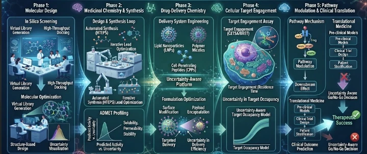
This article describes an uncertainty-aware platform for moving from predicted molecular binding to validated cellular target engagement in medicinal chemistry and drug-delivery research. Docking and machine-learning predictions can rapidly prioritize compounds, but they do not establish that a molecule reaches, occupies, and functionally modulates a target inside relevant cells. The article therefore separates predicted affinity from free intracellular concentration, subcellular localization, t
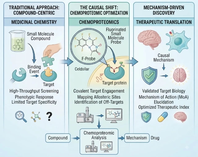
This article explores fluorinated medicinal chemistry through causal chemoproteomics, emphasizing that fluorine substitution can alter far more than potency. A single positional change can modify conformation, pKa, lipophilicity, metabolic stability, target interactions, intracellular exposure, and off-target biology. The article therefore tests whether fluorinated analogues change cellular phenotype through intended target engagement rather than hidden polypharmacology. Matched molecular pairs
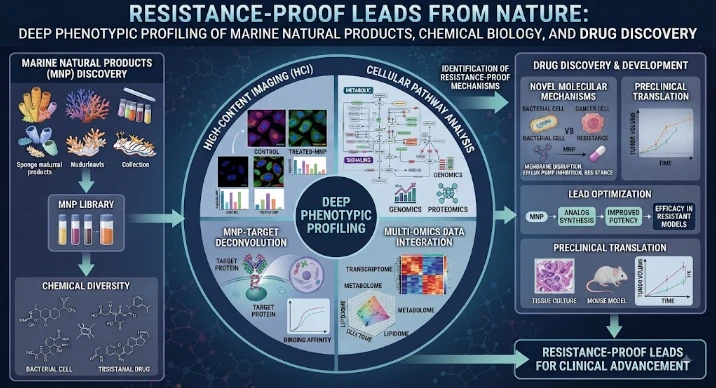
This article presents resistance-aware marine natural-product discovery as a mechanistic process rather than a search for isolated high-potency hits. It explains how activity from marine extracts or fractions can disappear during purification or fail under resistance pressure, making deep phenotypic profiling essential. Morphology, viability kinetics, organelle function, transcriptional state, metabolism, and recovery after washout are combined to connect phenotype with mechanism. The article em